British Heart Foundation: A colourful image of blood vessel cells wins competition
- Published
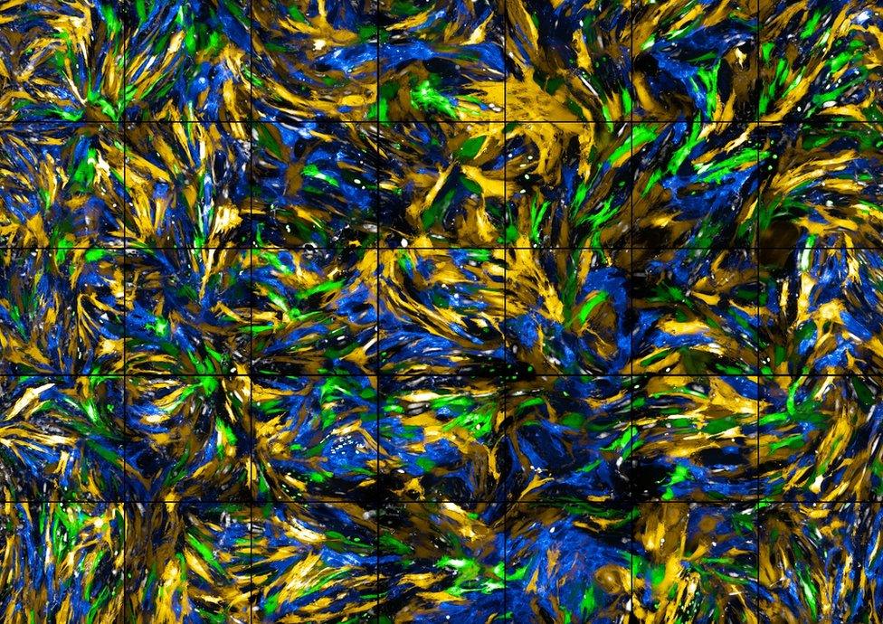
A colourful image of blood vessel cells has won this year's Reflections of Research competition, run by the British Heart Foundation (BHF).
The competition looks for the best images relating to research into heart and circulatory diseases, blending science and art.
The winning image, entitled A Sea of Cells (above), is a close-up of smooth muscle cells that line the blood vessels in mice, with the colours highlighting different proteins.
The researcher behind the image is Iona Cuthbertson, a PhD student at the University of Cambridge.
Ms Cuthbertson is exploring the ways in which rare types of smooth muscle cells in the walls of arteries grow rapidly after injury. Her research relates to conditions such as atherosclerosis, where there is a build-up of fatty substances inside arteries - a condition associated with increased stroke and heart attack risk.
"The winning image succeeds in portraying a turbulent drama happening at a cellular scale," said John O'Shea, guest judge and head of programming at Science Gallery London, King's College London.
"Through the skill and imagination of the scientists involved, all of the shortlisted images reveal to us in new ways [the] remarkable processes of life."
Here are the runners-up and shortlisted images, with descriptions provided by the BHF.

Judges' runner-up: The Forming Heart, by Dr Richard Tyser
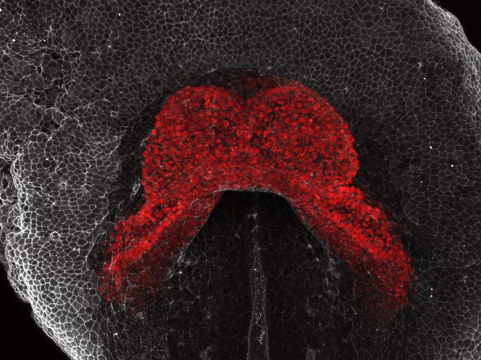
This image by Dr Richard Tyser, a BHF-funded research fellow at the University of Oxford, shows the heart in a developing mouse embryo.
The heart cells are coloured in red. During early development, the heart forms this crescent-like shape and starts to beat.

Supporters' favourite: The Heart and Brain Axis, by Cheryl Tan, Maryam Alsharqi, Dr Winok Lapidaire, Dr Mariane Bertagnolli and Dr Adam Lewandowski

BHF supporters chose the image above, which was a team effort by five researchers from the Radcliffe Department of Medicine at the University of Oxford.
The image reflects the interaction between the heart and the brain. The colours show the different imaging techniques, including: magnetic resonance imaging of the heart and brain, ultrasound imaging of the heart, and fluorescent imaging of the heart and blood vessels.
The following images were also shortlisted:

A Rush of Blood to the Head, by Dr Michael Drozd and Dr Nicole Watt, University of Leeds
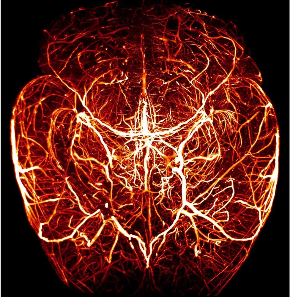
The complex network of blood vessels in the brain of a mouse.

Halo in the Heart, by Dr Alexander Fletcher and Dr Nicolas Spath, University of Edinburgh
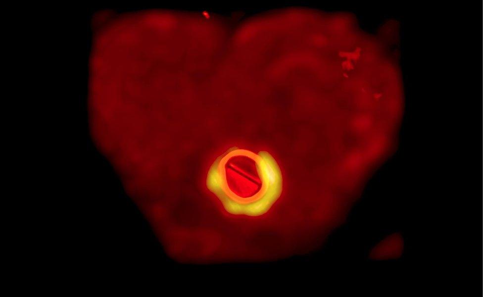
This is a scan of a heart viewed from below, with the bright "button" in the middle showing a metal valve that replaced the patient's natural heart valve.
The "halo" represents an area of high activity which suggests the new valve is infected.

A Blossom Pericyte, by Dr Elisa Avolio, University of Bristol
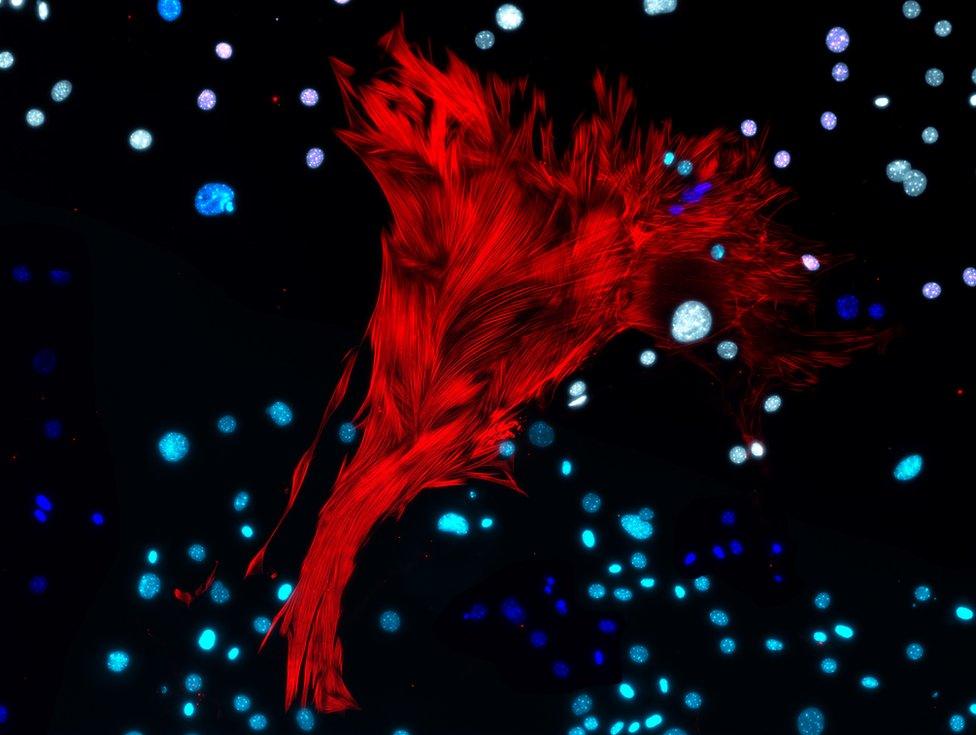
The image shows cells called pericytes which surround and support our blood vessels to make them stronger.

Blood-brain Barrier Rainbow, by Agne Stadulyte, University of Edinburgh

These colours represent different cells of the cerebellum within a mouse brain.
Astrocytes (green) are cells responsible for the relationship between neurons and blood vessels. TSPO (red) is a protein associated with steroid production.

Cutting off Life Support to the Heart, by Dr Mairi Brittan, University of Edinburgh
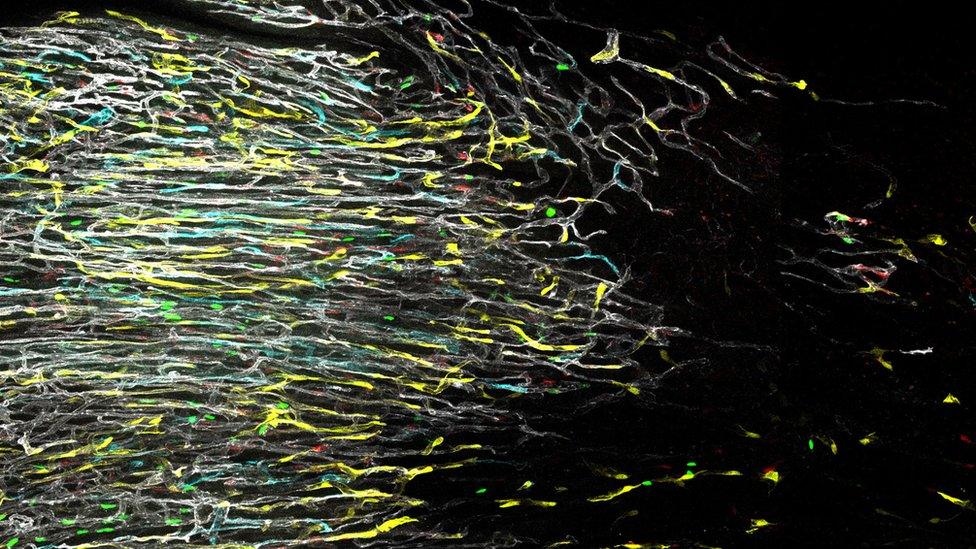
The image shows the blood vessel network in a mouse heart during the early stages after a heart attack.
The network of vessels comes to an abrupt stop at the edge of the injured heart muscle (left to right).

United Colours of Myocytes, by Dr Gabor Foldes, Dr Virpi Talman, Dr Harry Hartley, Imperial College London
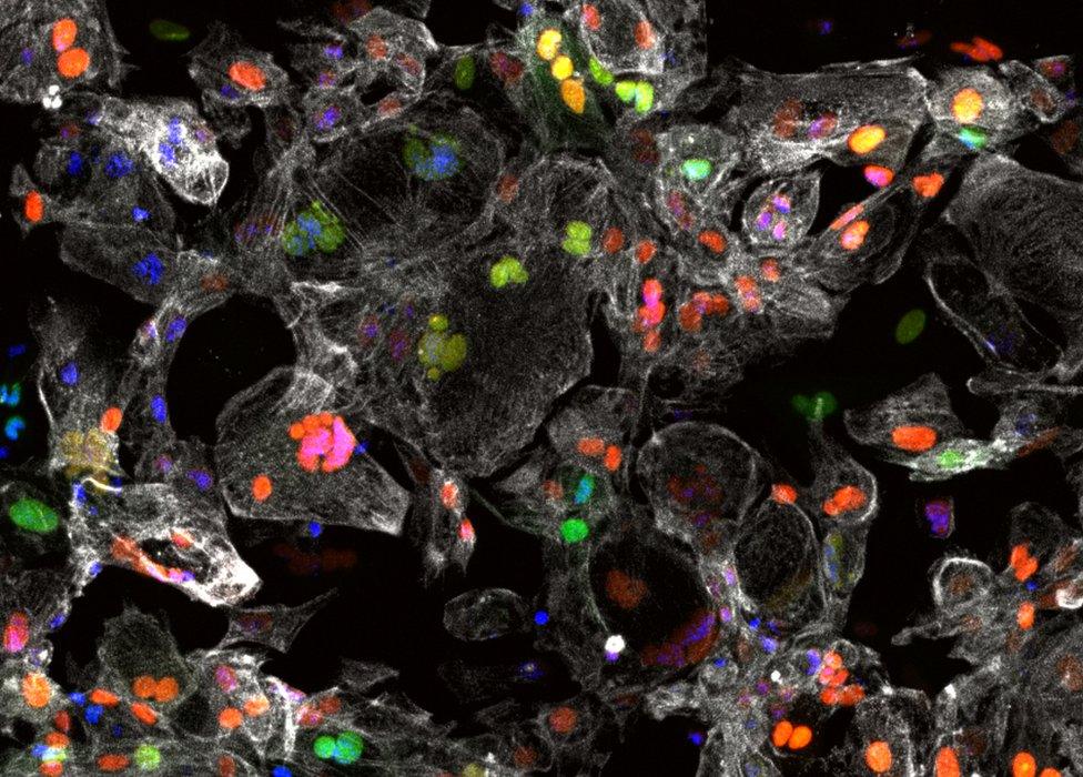
This image shows human heart muscle cells derived from stem cells. The different colours show a different phase of a cell's life cycle.
This colourful system is used to develop new treatments which can help the heart to renew itself after an injury, such as a heart attack.

Life-altering Algae, by Dr Ashish Patel, Dr Francesca Ludwinski, Prof Alberto Smith, Prof Suwan Jayasinghe and Prof Bijan Modarai, King's College London and University College London
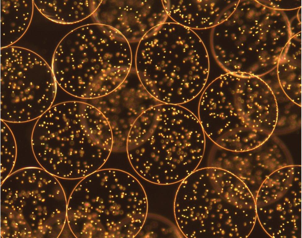
Here we have human monocytes, a type of white blood cell, that have been encapsulated in brown algae.
Injecting these capsules in the legs of people with severely limited blood flow may promote new blood vessel formation and restore blood flow to the damaged areas.
This algae-based treatment could reduce the need for amputations in people with critical limb ischaemia, when blood flow to the legs is severely restricted.

Nature's Bricks and Mortar, by Dr Fraser Macrae, University of Leeds
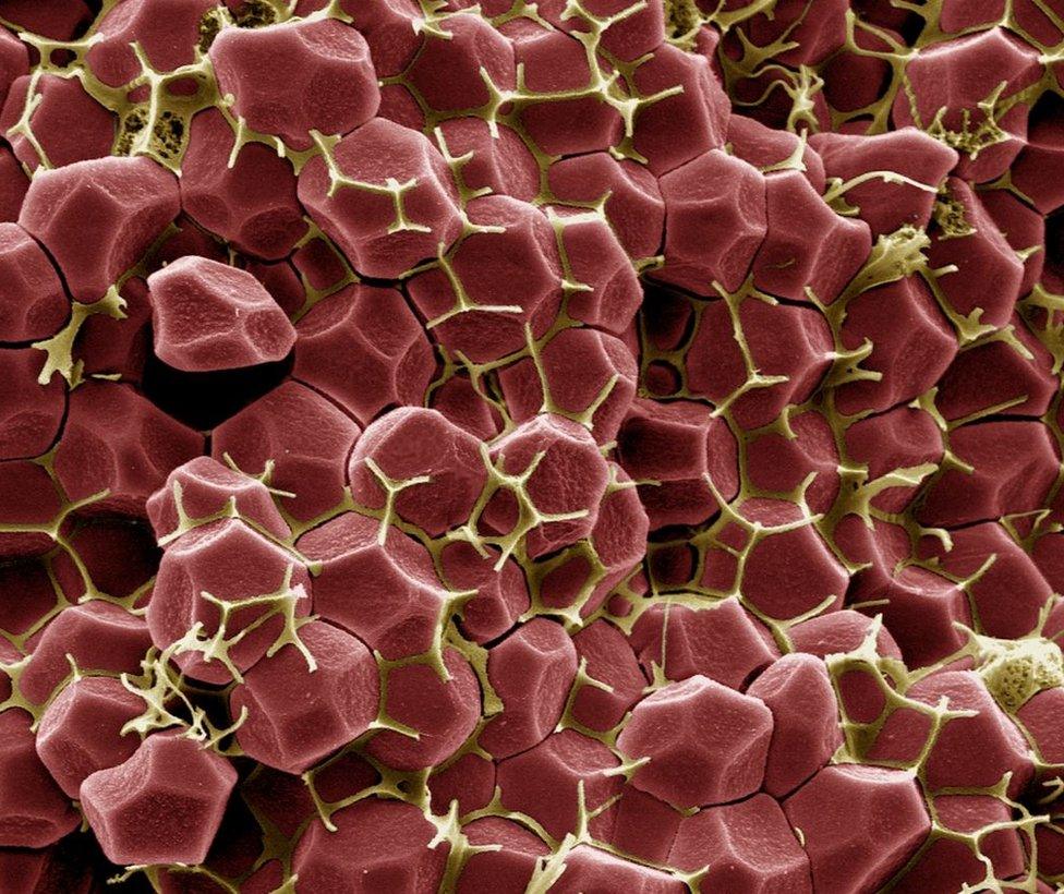
This image shows the internal structure of a blood clot. Blood clots contract, compressing red blood cells into polyhedral shapes forcing a protein called fibrin (yellow) into the gaps between them.
This creates an impermeable clot, perfect for preventing bleeding. But many cardiovascular diseases, like heart attacks and strokes are caused by the formation of obstructive blood clots in inconvenient places.
Images are copyright.
- Published10 July 2019

- Published8 February 2019
- Published11 January 2019