Hull Royal Infirmary uses X-ray guided surgery to remove blood clots
- Published

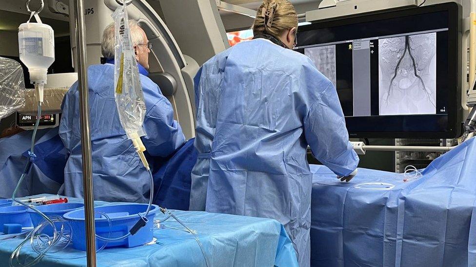
The vascular interventional radiology team at Hull Royal Infirmary use X-rays to guide their procedures
More potentially fatal blood clots are removed in Hull using X-ray guided technology than at any hospital in England, the trust has said.
The team at Hull Royal Infirmary operate on blood vessels and uncontrolled bleeding at the day unit.
About half of the operations performed each year are on emergency cases.
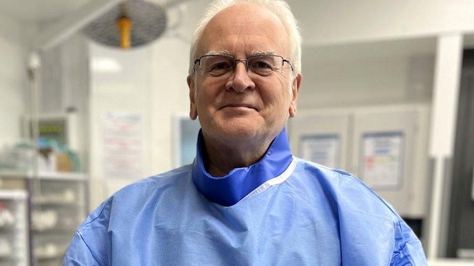
Prof Duncan Ettles said: "I think it's true to say we have more serious vascular disease here than anywhere else I have worked."
He said the X-ray guided technique is also used to carry out surgery on stroke patients to remove blood clots in the brain, operate on women with uncontrolled bleeding after childbirth and perform procedures on patients with cancer or kidney disease.
Prof Ettles, a consultant at Hull University Teaching Hospitals NHS Trust, said blood clots in the leg could "make walking excruciatingly painful and if left untreated, can end in amputation".

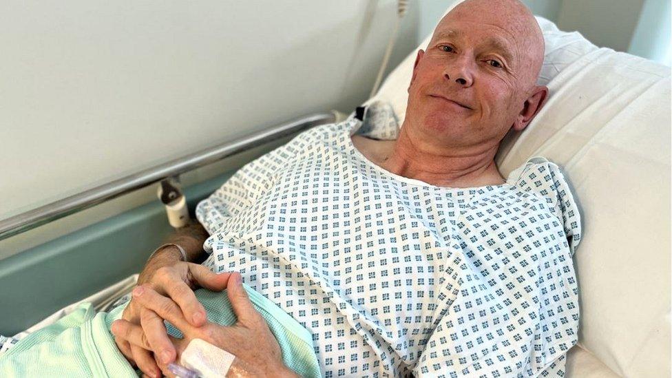
Keith Ritchie, 63, was able to go out for a walk with his dog within six hours of having the surgery
The vascular interventional radiology team at Hull Royal Infirmary have performed 1,122 vascular operations on patients from East Yorkshire and Northern Lincolnshire in the past year.
Between 2019 and 2021 they removed 1,161 clots from leg arteries, using X-ray guided surgery, which was around 10 times more than some hospital trusts, Prof Ettles said.
Keith Ritchie, 63, from Beverley, was operated on by Prof Ettles to remove a blood clot from an artery in his right leg.
Mr Ritchie, who works as a trainer at the Defence School of Transport in East Yorkshire, had started to experience pain in January.
"I could only walk about 100 metres and then had to stop to let the blood get down to the muscle," he said.
"It was quite frustrating and restricted a lot of things I like to do."

Prof Ettles said there was a link between high numbers of artery disease and communities with higher rates of deprivation
An angiogram, a type of X-ray which Prof Ettles described as creating "a roadmap of the blood vessels," was performed.
That was followed by an angioplasty operation, which involved feeding a wire, tube and balloon, through a tiny incision into the main artery of Mr Ritchie's leg and inflating it to clear the blockage.
The operation, carried out under local anaesthetic, took less than two hours.
"To be able to see the arteries opening up, that was really quite amazing. It looked easy to them, but really hard to me," Mr Ritchie said.
Mr Ritchie was back at home and able to take his dog Molly for a walk within six hours of leaving the operating theatre.
"What a colossal difference, I'm amazed actually. There's no pain whatsoever," he said.

Follow BBC East Yorkshire and Lincolnshire on Facebook, external, X (formerly Twitter), external, and Instagram, external. Send your story ideas to yorkslincs.news@bbc.co.uk, external