Human origins: 'Little Foot' fossil's big journey out of Africa
- Published
Looking inside the skull of "Little Foot" with X-rays causes no harm to the fossil
A priceless fossil was briefly brought to a UK research centre in complete secrecy two years ago, in an operation that had more than a touch of the spy novel about it.
The specimen was transported across South Africa with an armed guard, treated like an incognito VIP on an international flight, and then whisked slickly to the Diamond X-ray Light Source just south of Oxford.
It was at the British research facility that scientists were able to see some microscopic details in the ancient remains that could help unravel key clues to the origins of modern humans.
Details of the operation have been made public only now, as the first results from the X-ray investigations have been shared with the wider research community, external.
"It was immensely nerve-wracking," palaeoanthropologist Dominic Stratford recalls of the cloak-and-dagger mission, the first time any part of a prehistoric individual has been allowed out of South Africa.
Not only are the remains beyond value, after three million or more years embedded in sediments in the floor of a South African cave, they are immensely fragile.
What Prof Stratford had transported was the skull of "Little Foot", the most complete Australopithecine fossil ever recovered. And given the Australopithecines' position on the evolutionary road to modern humans, this makes Little Foot extra special.
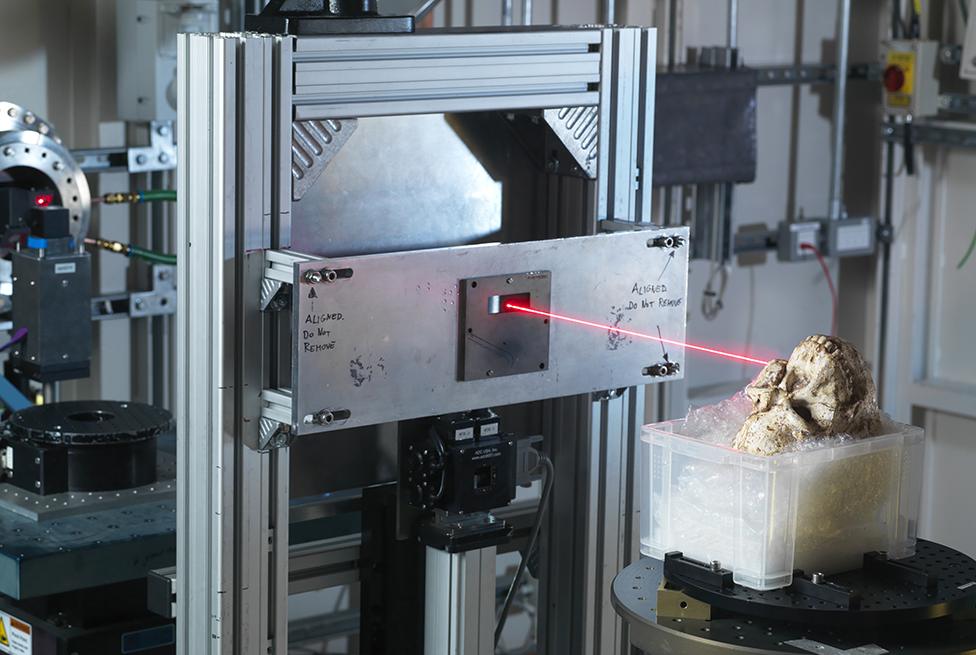
Penetrating, high-resolution X-rays get at hidden details without causing damage
Accompanying Prof Stratford and the skull was Ronald Clarke, the Witwatersrand University professor who led Little Foot's more-than-20-year excavation from the Sterkfontein Caves just outside Johannesburg.
Also in the party was Dr Amélie Beaudet, keen to use Diamond's powerful X-rays to peer inside the delicate object while doing no damage.
"With the X-rays, we found we could see tiny structures like the vasculature system, where there had been blood vessels inside Little Foot's bones, which normally would require physically slicing up a specimen," she told BBC News.
Prof Ian Tattersall, curator emeritus of human origins at the American Museum of Natural History in New York, is bowled over by the details revealed in the first scholarly paper to come out of the Diamond study.
"It is wonderful to have confirmation that micromorphology at this resolution can be recovered from a hominid this old," he said.
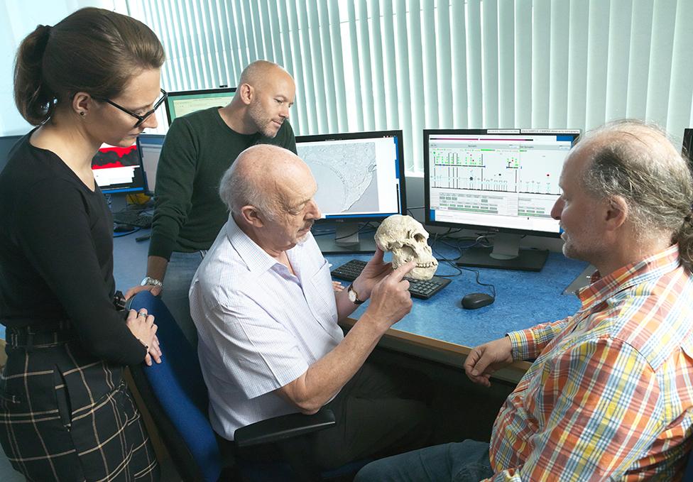
Ron Clarke has spent a large fraction of his career on this one specimen
From the Natural History Museum in London, Dr Louise Humphrey, a specialist in human origins and bioarchaeology not involved in the study, affirms the power of "this type of non-invasive investigation of microscopic structures… to reconstruct different aspects of an individual's life history from birth to death".
As an example, she highlights details in the tooth enamel revealed by the X-rays.
"Tooth enamel is not renewed during life," Dr Humphrey explains, "so it preserves a record of an individual's environment, diet and health during the first few years of life when the tooth crowns are developing."
Disrupted growth patterns revealed by the high-resolution images indicate "that [Little Foot] experienced at least two events that interrupted development during early life", she says.
Dr Beaudet told the BBC these tooth defects really stood out. "Dietary deficiency or nutritional stress" are blamed in the paper, though in an interview for BBC Radio 4's Inside Science programme, she accepted "we don't know if Little Foot was sick at some time, or if she couldn't find enough food".
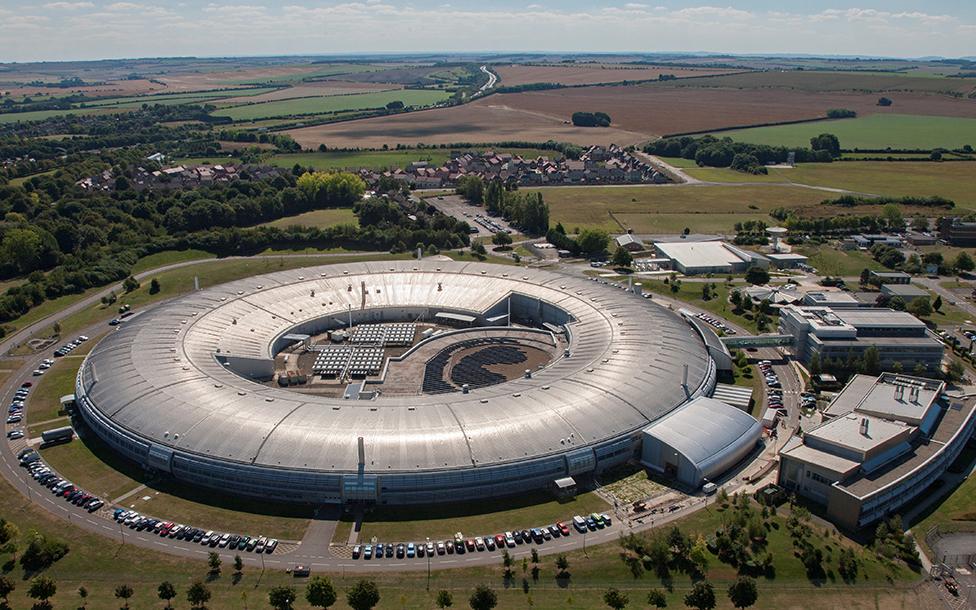
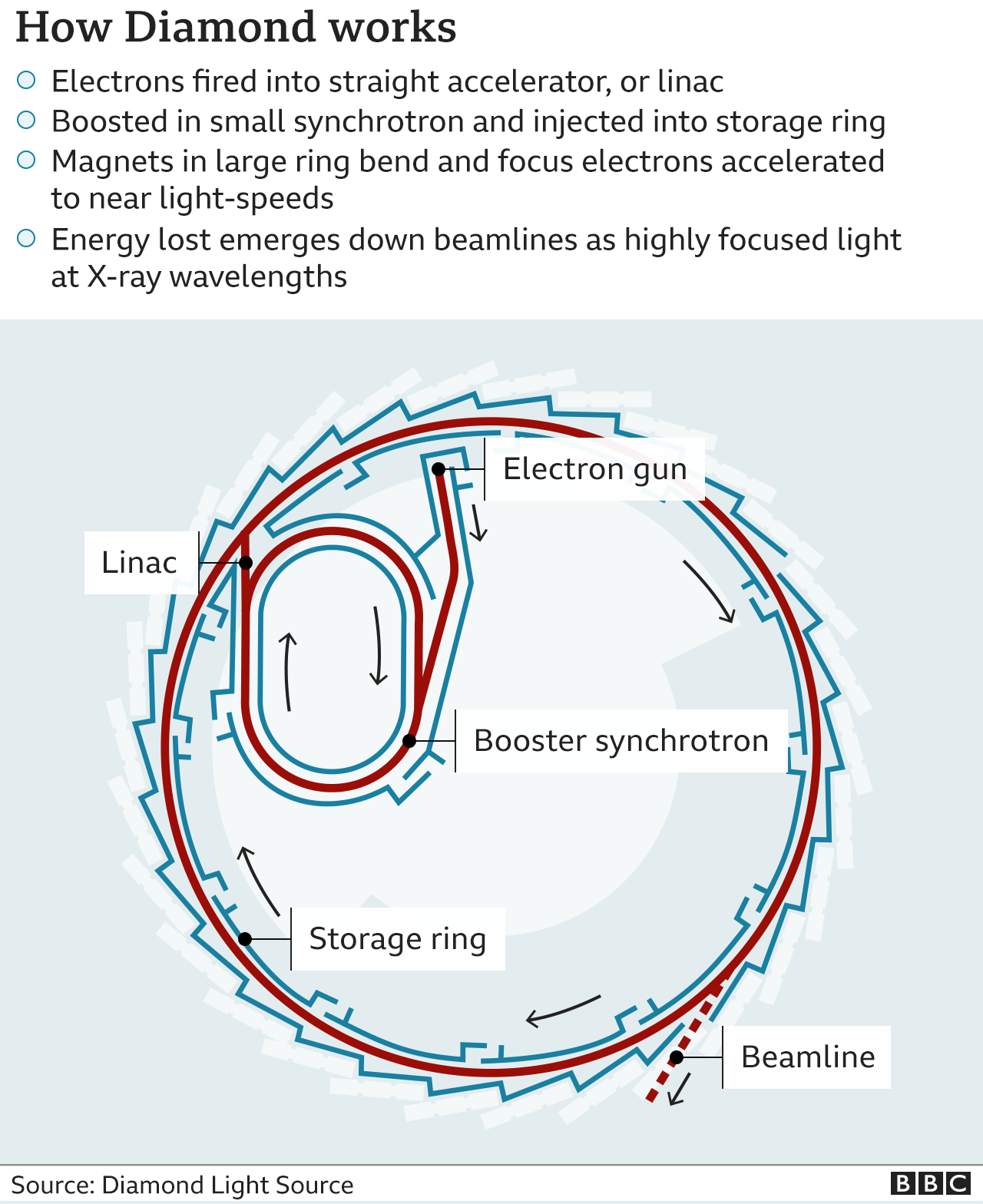
Diamond is what's known as a synchrotron. The giant machine produces brilliant X-rays
Dr Beaudet says they are also planning to measure a layer of material at the root of the teeth called cementum, which could indicate Little Foot's age when she died. It's thought death resulted when the creature fell into the cave through an opening in the ground.
The sheer amount of data the team managed to collect at Diamond, seven terabytes (140 Blu-ray discs), has been a challenge. Generating it was, too.
The skull, not much smaller than a modern human's, was far larger than they had been accustomed to examining at the synchrotron's "I12" imaging beamline.
The South African researchers had first sent a plastercast of the specimen, explained Thomas Connolley, the principal scientist on the beamline.
This meant the team could rehearse how best to mount the real object safely. And it was too large to be imaged all at once, so that new techniques had to be developed to knit together a patchwork of stills to create a complete 3D rendering - all the while conscious of the skull's importance in human prehistory.
"Only two people were allowed to handle the fossil," he reported. "Prof Clarke and Dominic Stratford."
He added: "None of us were allowed to touch it - with very good reason! Little Foot is probably the oldest and best-preserved fossils of this type.
"This was a very special experiment," Dr Connolley admitted. "It was actually quite emotional to think that we were studying one of our very early ancestors - for everyone I think, certainly for me."
Dominic Stratford confesses to a little emotion, too.
"It was a fantastic moment when we were all 'hutched' together in the beamline control room - it's always difficult to imagine what's preserved, so when we finally started to see some images, it was completely remarkable," he recalled.
Dr Beaudet, now based at Cambridge University after several years on Prof Clarke's team at Witwatersrand, says it is the imprint of the brain on the inside of the skull that will be most interesting for what it reveals of the early development of human intelligence.
Already the team has identified the traces of blood vessels in the inner skull that are similar to ones found in modern humans.
"The main hypothesis is that in modern humans, these vessels are involved in thermoregulation - preventing our brain from becoming too hot. With Little Foot, the brain was the same size as a chimpanzee's. It was only later in evolution that the brain grew dramatically. But at some point, something had to change in the vascular system, too. So the fact that we can see these vessels in Little Foot is quite promising," she said.
The problem is that at the moment, the team has no fossil comparisons - no study this detailed has been attempted before on ancient human remains.
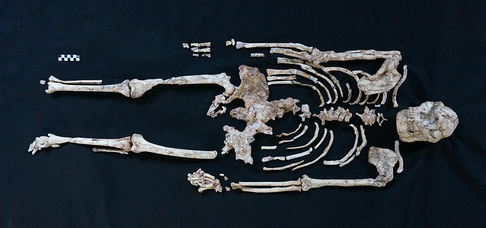
Little Foot is so nicknamed because of the diminutive nature of the bones of the feet
For palaeoanthropologist Ian Tattersall, this is what makes the preliminary data from Diamond so tantalising.
"While there are no amazing revelations here, this study opens large vistas for the future," he said. "And it is especially significant in demonstrating that micromorphology can be recovered from truly ancient hominid fossils without having to resort to destructive techniques. That is truly an exciting prospect."
There is much more work to be done on the skull scans already obtained, according to Dr Beaudet, but she is looking forward to examining the leg and arm bones, hands and feet of Little Foot, for what they will reveal of our ancestors' transition from tree-living to scurrying around on the ground.
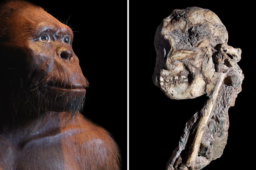
An artistic rendering of the Australopithecine (L) with part of the fossil remains (R), which were encased in rock. The fossil is thought to be about 3.6 million years old
The success with these first experiments reassures Prof Stratford.
"One of the blessings and curses of Little Foot is that we have these amazingly well-preserved, complete, single bones, which is almost unheard of. Fragments are easy to move around and to scan. But if you have a complete leg bone, or upper-arm and shoulder blade - that becomes a real challenge," he said.
"We have seen the potential to do this at Diamond. And that means we could reconstruct how Little Foot lived on the landscape, how she moved around, what kind of stresses she was putting the bones under. And we could fit that into the big picture of human evolution at the time."